
Occult Scaphoid Fracture: Why Your X-ray Was Normal
Up to 20% of scaphoid fractures are missed on initial X-ray. MRI within 2 weeks rules out occult fracture and prevents non-union — when to ask for follow-up imaging.

A fall on an outstretched hand — known clinically as a FOOSH injury — is one of the most common mechanisms behind scaphoid fractures. Yet 15 to 20% of true scaphoid fractures produce a completely normal initial wrist X-ray. Walking away from the emergency department with a 'normal' X-ray does not rule out a fracture, and returning to activity without proper immobilization can lead to non-union — a situation where the fracture never heals — and eventually post-traumatic arthritis known as SNAC wrist (scaphoid non-union advanced collapse).
The standard of care when clinical suspicion remains high despite a negative X-ray is either repeat imaging in 10 to 14 days or, ideally, MRI within 7 days. Understanding why your X-ray was normal, which clinical signs should prompt follow-up, and why MRI is the superior next step can help you advocate for the right care before permanent damage occurs.
Why Scaphoid Fractures Hide on X-ray
The scaphoid is a small, boat-shaped carpal bone with a complex three-dimensional anatomy. Non-displaced waist fractures — the most common type — run through the narrowest part of the bone at an oblique angle. On a standard PA wrist X-ray, the fracture line is obscured by the overlapping shadows of surrounding carpal bones, and because the fracture is not displaced, there is no step or gap to catch the eye. The fracture line can be as thin as a hair's breadth on the first day, well below the resolution of plain radiography.
Additionally, the initial X-ray is taken within hours of injury when soft-tissue swelling and bone resorption along the fracture margins — the changes that eventually make the fracture visible — have not yet developed. Repeat X-rays taken 10 to 14 days later may reveal the fracture as the edges resorb, but by then valuable healing time has passed if the wrist was not immobilized.
Clinical Signs That Should Trigger Follow-up Imaging
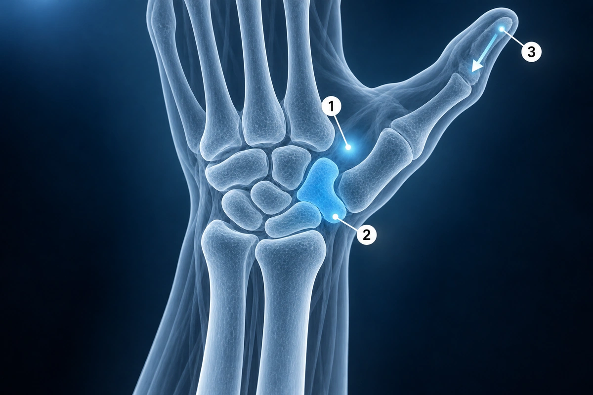
If you have any of the following after a fall on an outstretched hand, insist on follow-up imaging even if your X-ray was reported as normal:
- Anatomical snuffbox tenderness — pain in the hollow on the back of the wrist between the thumb tendons when pressed
- Scaphoid tubercle tenderness — pain at the bony prominence on the palm side of the wrist below the thumb
- Pain with axial compression of the thumb — pressing the tip of the thumb toward the wrist reproduces pain
- Pain with radial deviation — moving the wrist toward the thumb side hurts
- FOOSH mechanism with persistent pain beyond 48 hours — pain that does not improve rapidly is a red flag
Anatomical snuffbox tenderness alone has a sensitivity of around 90% for scaphoid fracture — meaning it is present in nearly all confirmed fractures. Its specificity is lower, so a positive test does not prove a fracture, but a negative test in a calm patient is reassuring.
The Standard Protocol When X-ray Is Normal but Pain Persists
Current evidence-based guidelines recommend one of three pathways when clinical suspicion for occult scaphoid fracture is present despite a normal X-ray:
- Immobilize in a thumb spica splint immediately and repeat X-ray in 10 to 14 days — acceptable when MRI is not readily available and clinical suspicion is moderate
- MRI within 7 days — the preferred path when clinical suspicion is high; bone marrow edema appears within hours of fracture and is detectable on T2 fat-saturated sequences long before the fracture line becomes visible on X-ray
- CT scan — useful for high-energy mechanisms or when MRI is contraindicated; CT detects fracture lines but misses bone bruise and cannot exclude a fracture without one
During the waiting period for any of these pathways, the wrist must be immobilized in a thumb spica splint. Activity that loads the scaphoid — gripping, weightbearing through the wrist, racket sports, gymnastics — risks displacing an undisplaced fracture and converting a simple cast case into one requiring surgery.
Why MRI Beats Repeat X-ray
MRI has a sensitivity of approximately 98 to 100% for occult scaphoid fractures, compared to roughly 70% for a repeat X-ray taken at two weeks. The key difference is what each modality detects. X-ray requires a visible fracture line or bone resorption. MRI detects bone marrow edema — the flooding of the bone with fluid that occurs within hours of any significant injury — as bright signal on T2 fat-saturated or STIR sequences. This edema is present even when there is only a bone bruise (trabecular microfracture) without a complete fracture line.
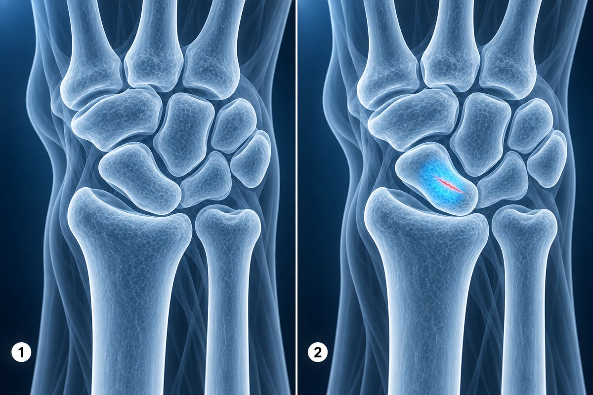
A normal MRI rules out both a fracture and a significant bone bruise, allowing you to return to activity immediately and avoid weeks of unnecessary immobilization. A two-week repeat X-ray cannot offer this — even if negative, clinical uncertainty remains. For a deeper understanding of what wrist MRI reveals, see our guide on how to read wrist MRI.
Cost-Benefit: One MRI vs. a Lifetime of Consequences
A wrist MRI costs several hundred to a few thousand dollars depending on your location and insurance. A missed scaphoid fracture that progresses to non-union requires bone grafting surgery. If the non-union goes untreated, it leads to SNAC wrist — a predictable cascade of carpal collapse and arthritis that ultimately requires salvage procedures including partial or total wrist fusion. The total cost, disability, and quality-of-life impact of SNAC wrist far exceeds the cost of a timely MRI.
Several health economic analyses have confirmed that early MRI — even in a lower-suspicion population — is cost-effective compared to a 'splint and repeat X-ray' strategy when the costs of missed fractures are modeled over a lifetime horizon. If your insurer or doctor hesitates, this evidence can support your request.
Red Flags for Urgent Imaging
Certain features should prompt urgent same-day or next-day imaging rather than a two-week wait:
- Proximal pole tenderness — the proximal pole of the scaphoid has poor blood supply and a much higher rate of non-union and avascular necrosis; any fracture here requires early diagnosis and often surgery
- Visible deformity or swelling out of proportion to the reported mechanism
- High-energy mechanism such as a motorcycle or sports collision rather than a simple domestic fall
- Occupation or sport that cannot tolerate prolonged immobilization — early surgery for a confirmed fracture returns these patients faster than weeks in a cast
For patients whose fractures are confirmed, treatment decisions depend heavily on fracture location and displacement. Our article on whether a scaphoid fracture can heal without surgery covers those decisions in detail. For AI analysis of your wrist imaging, visit the scaphoid fracture condition page.
Key Takeaways
- 15 to 20% of true scaphoid fractures are invisible on the initial wrist X-ray — a normal X-ray does not rule out a fracture
- Anatomical snuffbox tenderness, scaphoid tubercle tenderness, and axial thumb compression pain after a FOOSH all warrant follow-up imaging despite a negative X-ray
- Immobilize in a thumb spica splint immediately while awaiting definitive imaging — do not return to activity with persistent wrist pain after a fall
- MRI within 7 days is the gold standard: sensitivity approaches 100% by detecting bone marrow edema that appears within hours of injury
- A normal MRI clears you to resume activity immediately; repeat X-ray at two weeks cannot provide that certainty
- Proximal pole tenderness, high-energy mechanism, and visible deformity are red flags for urgent imaging rather than a two-week wait
Frequently Asked Questions
How soon should I get the MRI after injury?
Within 7 days is ideal. Bone marrow edema is detectable on MRI within hours of significant wrist trauma, so there is no benefit in waiting. Earlier imaging also means earlier definitive treatment — confirmed fractures can be immobilized or scheduled for surgery without losing additional weeks. If you cannot access MRI within 7 days, apply a thumb spica splint immediately and arrange the scan as soon as possible.
My doctor sent me home with a normal X-ray and pain that persists — what should I do?
Contact your doctor and specifically request either an MRI or a repeat X-ray at two weeks. In the meantime, buy or borrow a thumb spica splint (widely available at pharmacies) and avoid any loading of the wrist. Explain your symptoms clearly: tenderness in the anatomical snuffbox, pain with thumb compression, and mechanism of a fall on an outstretched hand. These three together constitute high clinical suspicion and should prompt follow-up imaging under current guidelines. If your GP is reluctant, an urgent orthopaedic or hand surgery consultation is appropriate.
Will MRI show a non-union if my fracture was already missed?
Yes. MRI is excellent at demonstrating established non-union. A non-union appears as a persistent low-signal fracture line on T1, often with cystic change along the margins and a humpback deformity of the scaphoid. MRI also shows avascular necrosis of the proximal pole as diffusely low T1 signal without contrast enhancement, which is critical for surgical planning. CT is often added for precise measurement of the gap and angulation when surgery is planned.
Is the MRI cost worth it compared to waiting for a repeat X-ray?
For most patients with persistent symptoms and a plausible mechanism, yes. A timely MRI costs a fraction of the bone grafting surgery needed to treat established non-union, and a vanishingly small fraction of the lifetime cost of SNAC wrist arthritis and salvage procedures. If a normal MRI clears you to return to work and activity immediately, it also saves weeks of lost productivity and avoids the long-term wrist stiffness that follows unnecessary immobilization.
What happens if a scaphoid fracture goes completely untreated?
Without treatment, a missed scaphoid fracture progresses to non-union in a significant proportion of cases — the exact rate depends on fracture location, but proximal pole fractures approach 30% non-union even with treatment. Established non-union leads to progressive carpal collapse as the scaphoid rotates and loses height. This produces the SNAC wrist pattern: a predictable sequence of arthritis affecting the radioscaphoid joint first, then the capitolunate joint. Eventually the entire wrist joint is destroyed. By the time SNAC wrist is symptomatic enough to seek care, the only options are partial or total wrist fusion — interventions that permanently limit wrist movement. Early diagnosis and treatment, by contrast, gives the majority of patients a fully functional wrist.
Related Articles
Understand scaphoid fracture healing rates by location, cast treatment versus surgical fixation, and the blood supply challenge.
Decode your wrist MRI report including TFCC evaluation, scaphoid fracture detection, and carpal tunnel findings.
Learn about common wrist conditions including TFCC tears, scaphoid fractures, carpal tunnel syndrome, de Quervain's, and ganglion cysts.
Related Conditions
Ready to analyze your imaging? Upload your MRI or X-ray for AI-powered analysis
Upload your MRI or X-ray DICOM files for private, AI-powered analysis. 4 models analyze independently — raw scan files stay in your browser.
Start AnalysisMedical Disclaimer: This page is for informational and educational purposes only. It does not constitute medical advice, diagnosis, or treatment. AI-generated analysis may contain errors. Always consult a qualified healthcare professional for medical decisions. Full Disclaimer