
Common Symptoms
- Chronic ulnar-sided wrist pain, typically gradual in onset
- Pain worse with grip, ulnar deviation, and forearm pronation
- Painful clicking or clunking with forearm rotation
- Weakness and reduced grip strength
- Gradual onset without a specific injury event
- History of distal radius fracture or congenital positive ulnar variance
Ulnar Impaction Syndrome on MRI: AI-Powered Imaging Analysis
AI-powered ulnar impaction (abutment) syndrome analysis on wrist MRI. Understand positive ulnar variance, lunate bone marrow edema, TFCC degeneration, and imaging findings in plain language.
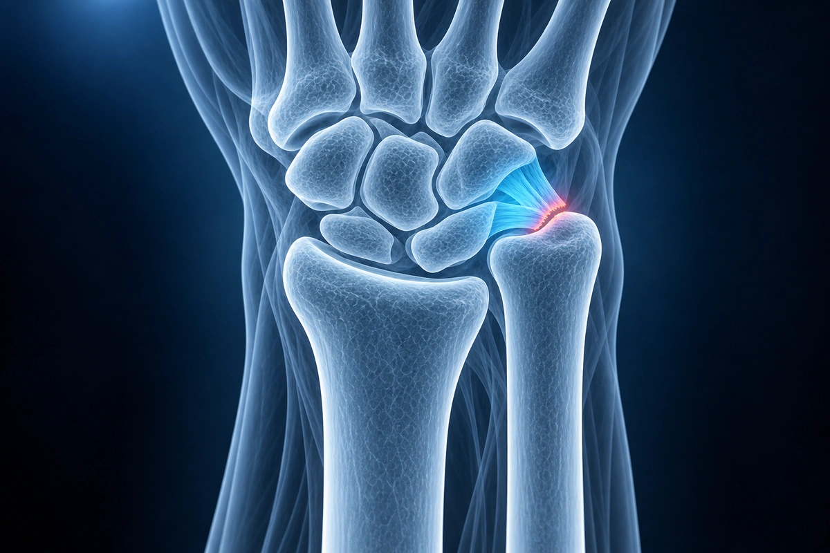
Ulnar impaction syndrome — also called ulnar abutment syndrome or ulnocarpal abutment — occurs when the ulna is relatively longer than the radius (positive ulnar variance), causing it to repetitively load the ulnar side of the wrist during grip and forearm rotation. The structures that absorb this excess load are the lunate, the triquetrum, and the triangular fibrocartilage complex (TFCC). Over months to years, this chronic mechanical stress produces a predictable sequence of injury: cartilage wear on the ulnar head and the lunate facet, bone marrow edema and subchondral cysts in the lunate and triquetrum, central perforation of the TFCC, and eventually tearing of the lunotriquetral ligament. Treatment focuses on reducing the load on the ulnar side of the wrist, either through activity modification and splinting or, when conservative measures fail, by surgically shortening the ulna.
Key Imaging Findings
- Positive ulnar variance on a neutral posteroanterior wrist X-ray — the ulnar articular surface sits more than 2 mm distal to the radial articular surface
- Lunate bone marrow edema on T2 fat-saturated MRI at the ulnar and proximal aspect of the lunate — the site of greatest ulnar contact stress
- Subchondral cyst formation or edema in the triquetrum at the lunotriquetral articulation
- Central TFCC perforation (Palmer class 2A through 2E degenerative spectrum) — seen as a full-thickness signal defect through the disc on T2 or PD fat-sat sequences
- Lunotriquetral ligament tear in advanced cases — partial or complete disruption of the membranous or dorsal component
- Ulnar head chondral wear and corresponding lunate facet chondral loss on high-resolution MRI or MR arthrography
Frequently Asked Questions
Do I need surgery for ulnar impaction syndrome?
Not necessarily. The first-line approach is a 3 to 6 month trial of conservative management: activity modification to avoid loading the ulnar wrist, a neutral or slight radial-deviation wrist splint for symptom-provoking tasks, anti-inflammatory medication, and physiotherapy to reduce pronation loading and strengthen the forearm. Corticosteroid injection into the distal radioulnar joint or ulnocarpal space can provide medium-term relief and help confirm the diagnosis. If symptoms persist beyond 6 months of consistent conservative care, or if imaging shows progressive lunate changes, ulnar shortening osteotomy is the definitive surgical solution. The osteotomy removes a measured segment of the ulnar shaft to restore neutral variance, reducing ulnocarpal contact forces by over 40%. Arthroscopic wafer resection — removing the distal few millimeters of the ulnar head through the wrist — is an alternative for mild positive variance when open surgery is not preferred.
Can a wrist splint help ulnar impaction syndrome?
Yes, for managing symptoms, though not for correcting the underlying anatomy. A wrist splint worn during activities that provoke pain — particularly tasks requiring grip with the forearm in pronation or ulnar deviation — reduces the mechanical load transmitted through the ulnocarpal joint. Neutral or slight radial deviation positioning decreases contact between the ulnar head and the lunate. Splinting is most useful during the conservative treatment phase and for protecting the wrist after ulnar shortening surgery while the osteotomy heals.
How is ulnar variance measured and what counts as positive?
Ulnar variance is measured on a standardized posteroanterior wrist X-ray taken with the shoulder abducted 90 degrees, elbow flexed 90 degrees, and the forearm in neutral rotation — this is critical because forearm pronation artificially increases apparent ulnar variance by up to 2 mm, and supination decreases it. On this neutral PA view, the distance between the line of the radial articular surface and the line of the ulnar articular surface is measured. Neutral variance means the two surfaces are at the same level. Positive ulnar variance means the ulna projects more distally than the radius; a variance greater than 2 mm is considered clinically significant and correlates with a substantially increased risk of ulnar impaction syndrome, central TFCC perforation, and lunate chondral changes.
Upload your imaging to check for Ulnar Impaction
Upload your MRI or X-ray DICOM files for private, AI-powered analysis. 4 models analyze independently — raw scan files stay in your browser.
Upload & AnalyzeMedical Disclaimer: This page is for informational and educational purposes only. It does not constitute medical advice, diagnosis, or treatment. AI-generated analysis may contain errors. Always consult a qualified healthcare professional for medical decisions. Full Disclaimer