
Kienbock Disease: Lichtman Stages Explained on MRI
Lichtman stages I–IV of Kienbock disease — lunate avascular necrosis, MRI signal evolution, fragmentation, collapse, and treatment by stage.
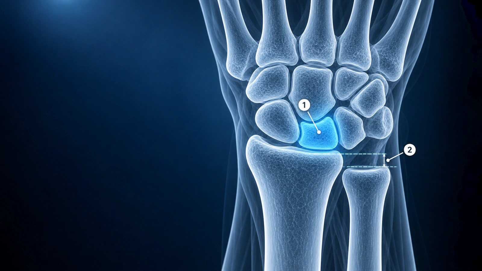
Kienbock's disease is avascular necrosis of the lunate — the central keystone bone of the proximal carpal row. The lunate's tenuous vascular supply makes it susceptible to ischemic injury from repetitive compressive loading, vascular anatomic variants with limited collateral flow, or ulnar minus variance, a skeletal alignment in which the ulna is shorter than the radius. This anatomy concentrates abnormal load on the radiolunate joint. The exact pathway from these risk factors to osteonecrosis remains incompletely understood, but the clinical result is predictable progressive lunate deterioration when left untreated.
Staging Kienbock's disease is essential for treatment planning because the optimal intervention differs dramatically across disease progression. The Lichtman classification — refined by the Stahl modification, which adds Stage IIIC for coronal split fractures of the lunate — is the most widely adopted system. It tracks the radiographic and MRI evolution from normal X-ray with marrow edema only, through sclerosis, fragmentation, carpal collapse, and eventual pancarpal arthritis. MRI is the cornerstone of diagnosis and staging because it detects Stage I disease weeks to months before any radiographic abnormality is visible.
Stage I: Normal X-ray, MRI Detects Edema
In Stage I, plain radiographs are entirely normal — no sclerosis, no height loss, no cortical irregularity. The lunate appears radiographically indistinguishable from a healthy bone. MRI reveals the diagnosis: diffuse hypointensity throughout the lunate on T1-weighted sequences, reflecting replacement of normal fatty marrow by ischemic or edematous tissue. T2-weighted signal is variable and depends on the relative contribution of acute edema versus early fibrous replacement. When acute edema dominates, T2 signal is elevated; as ischemia progresses toward sclerosis, T2 signal normalizes or decreases.
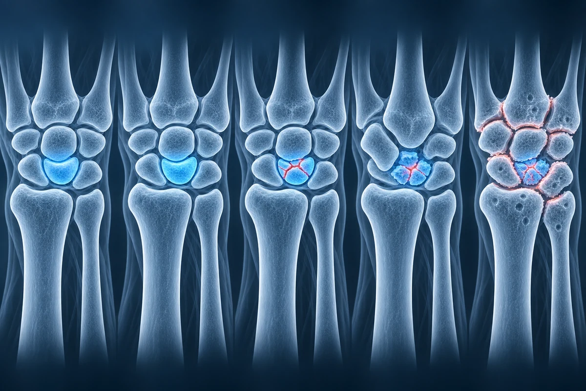
Stage I is the therapeutically critical window. The lunate retains its structural integrity, so interventions aimed at restoring vascular supply or reducing mechanical load have the greatest chance of halting progression. Gadolinium-enhanced MRI can demonstrate enhancement in viable portions of the lunate, guiding revascularization planning. For guidance on interpreting wrist MRI sequences, see our article on how to read a wrist MRI.
Stage II: Sclerosis Without Collapse
Stage II Kienbock's disease is defined by the appearance of lunate sclerosis on plain radiograph — a diffuse or patchy increase in bone density reflecting dead bone and reactive mineralization — without any loss of lunate height or carpal architectural change. The lunate contour remains intact. On MRI, T1 signal remains diffusely low. T2 signal in Stage II is typically low as well because edema has given way to sclerotic dead bone with little free water; this distinguishes Stage II from Stage I at the MRI level even when radiographs show only early sclerosis.
Stage II still offers an intervention window before collapse. Joint leveling procedures — radial shortening osteotomy or ulnar lengthening osteotomy — are most commonly offered in Stage I and II when ulnar minus variance is present, because unloading the radiolunate joint can slow or arrest progression. Revascularization with a vascularized bone graft may also be considered in Stage II.
Stage IIIA: Fragmentation Without Carpal Collapse
Stage IIIA marks the beginning of structural failure. The lunate shows collapse, fragmentation, or a coronal fracture line on radiograph or CT. However, overall carpal alignment is preserved: the scaphoid retains normal orientation, the capitate has not migrated proximally, and the carpal height ratio remains within normal limits. The Stahl-modified classification identifies a subgroup — Stage IIIC — for lunates with a frank coronal split fracture, which carries a distinct surgical implication of potential vascularized distal radius bone graft insertion into the fracture plane.
With carpal alignment still intact, motion-preserving procedures remain viable in Stage IIIA. Capitate shortening osteotomy, which unloads the lunate from the distal row, and vascularized bone grafts from the dorsal distal radius are preferred options. Because the carpus has not yet collapsed, these procedures aim to preserve functional wrist motion alongside arresting disease progression.
Stage IIIB: Fragmentation With Carpal Collapse
Stage IIIB represents a qualitative shift in disease severity. Lunate fragmentation is accompanied by fixed carpal collapse: the scaphoid assumes a flexed, rotated posture (the scaphoid ring sign on PA radiograph), and the capitate migrates proximally through the space vacated by the collapsed lunate. This carpal malalignment pattern resembles DISI (dorsal intercalated segment instability) and is not reducible by traction. Carpal height ratio is reduced. The radiocarpal joint may still be spared at this stage.
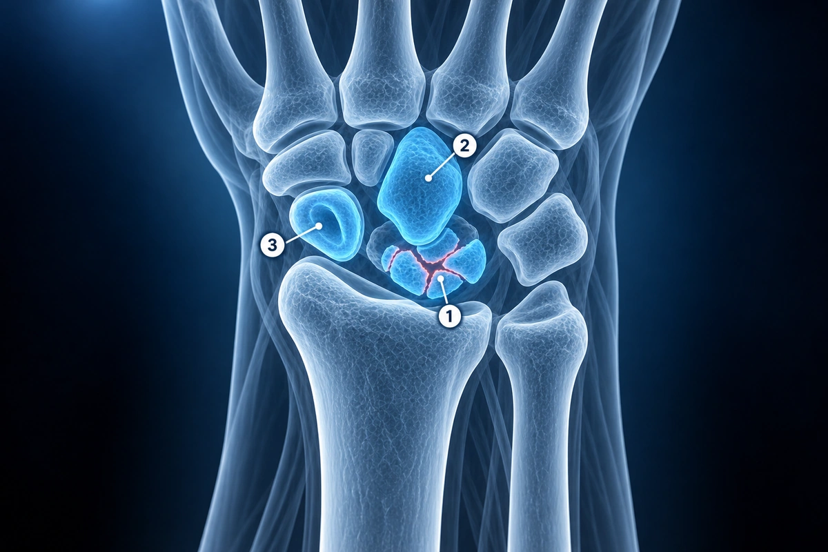
The irreversibility of carpal collapse in Stage IIIB shifts treatment toward salvage procedures that accept limited motion in exchange for pain relief and stability. Proximal row carpectomy removes the scaphoid, lunate, and triquetrum entirely, creating a new articulation between the capitate and the lunate fossa of the radius — suitable when the capitate head cartilage and lunate fossa are preserved. Scaphotrapeziotrapezoid (STT) fusion or four-corner fusion (capitate-hamate-triquetrum-lunate) corrects carpal alignment while eliminating motion at the midcarpal joint. See our overview of Kienbock's disease for the full clinical picture.
Stage IV: Pancarpal Arthritis
Stage IV is the end-stage of Kienbock's disease. The collapsed, fragmented lunate has driven generalized articular cartilage loss across both the radiocarpal and midcarpal joints. Radiographs show diffuse joint space narrowing, osteophyte formation, and subchondral cyst formation throughout the wrist. MRI demonstrates low T1 and T2 signal throughout the carpus with articular cartilage loss evident on fat-suppressed sequences. The clinical presentation is chronic, fixed wrist pain with severely limited range of motion and grip strength.
At Stage IV, the only reliable pain-relieving options are total wrist arthrodesis (fusion of the radiocarpal and midcarpal joints, sacrificing all wrist motion but providing a stable, pain-free extremity for grip-dependent tasks) or total wrist arthroplasty (a prosthetic joint replacement that preserves some functional motion but carries a higher reoperation rate). Patient factors — age, activity demands, and hand dominance — drive the final decision between these two options.
Treatment by Stage
Stage I treatment focuses on offloading the lunate and maximizing revascularization potential. Immobilization with a short-arm cast for 3-6 months is first-line in lower-demand patients or when surgery is contraindicated. In patients with ulnar minus variance, radial shortening osteotomy is the most studied joint leveling procedure, reducing radiolunate contact stress and stabilizing the TFCC. Vascularized bone grafts from the pronator quadratus or the dorsal distal radius (4th and 5th extensor compartment artery graft) deliver a new vascular pedicle into the ischemic lunate.
Stage II mirrors Stage I in terms of joint leveling eligibility. The key distinction from Stage IIIA is the absence of lunate structural failure, which means the joint leveling effect can still benefit an architecturally intact lunate. Stage IIIA adds capitate shortening osteotomy as a surgical option — reducing the length of the capitate unloads the lunate from below without requiring a formal joint leveling of the forearm bones. Vascularized bone grafts remain viable in well-selected Stage IIIA patients before carpal collapse becomes fixed.
Stage IIIB management pivots to salvage: proximal row carpectomy preserves grip-range wrist motion when capitate cartilage is intact; four-corner fusion sacrifices midcarpal motion but provides better stability when the capitate head shows articular damage. Stage IV leaves total wrist arthrodesis or arthroplasty as the two remaining options, chosen based on patient age, demand, and tolerance of zero versus limited residual motion.
Key Takeaways
- MRI is the only modality that detects Stage I Kienbock's disease — diffuse T1 hypointensity with normal radiographs defines this critical early window
- Lichtman staging runs I → II (sclerosis) → IIIA (collapse, aligned carpus) → IIIB (collapse, carpal malalignment) → IV (pancarpal arthritis); Stahl adds IIIC for coronal split fractures
- Ulnar minus variance concentrates load on the radiolunate joint and is present in ~75% of Kienbock's patients; joint leveling (radial shortening or ulnar lengthening) is the primary Stage I-II intervention when variance is present
- Stage IIIB carpal collapse — scaphoid flexion and proximal capitate migration — is fixed and irreversible, shifting treatment to salvage procedures such as proximal row carpectomy or four-corner fusion
- Stage IV pancarpal arthritis is managed by total wrist arthrodesis or arthroplasty; patient age and functional demands determine which option is appropriate
- Gadolinium-enhanced MRI can demonstrate residual lunate vascularity, guiding revascularization candidacy in Stages I and II
Frequently Asked Questions
Why is MRI preferred over X-ray and CT for diagnosing early Kienbock's disease?
Stage I Kienbock's disease produces no visible abnormality on plain radiographs or CT because the lunate retains its shape and density before structural failure begins. MRI detects bone marrow ischemia through signal changes in the fat-containing marrow: diffuse T1 hypointensity indicates marrow replacement by necrotic and edematous tissue weeks to months before radiographic sclerosis appears. This early detection window matters clinically because Stage I and II interventions — joint leveling, revascularization — have significantly better outcomes than treatment initiated after Stage IIIA collapse. CT is complementary for characterizing coronal fracture lines and planning surgery once structural failure is already present on radiograph.
What is ulnar minus variance and why does it matter for Kienbock's disease?
Ulnar variance describes the length relationship between the distal ulna and the distal radius measured on a standardized PA radiograph with the forearm in neutral rotation. Ulnar minus variance means the ulna is shorter than the radius — typically defined as more than 1 mm shorter. This alignment reduces the force-sharing contribution of the triangular fibrocartilage complex (TFCC) and concentrates a disproportionate share of axial forearm load onto the radiolunate articulation. Studies show ulnar minus variance in approximately 75% of Kienbock's patients compared to 20% of the general population, implicating it as a significant biomechanical risk factor. Joint leveling procedures — radial shortening osteotomy or ulnar lengthening osteotomy — correct this asymmetry, redistributing load and slowing or halting lunate ischemia in Stages I and II.
Can Kienbock's disease reverse or stabilize on its own?
Spontaneous revascularization and symptom stabilization have been reported in Stage I and early Stage II, particularly in patients who undergo prolonged immobilization and activity modification. However, Kienbock's disease does not reliably self-resolve, and untreated progression from Stage I to Stage IIIA has been documented over periods of months to years. Once Stage IIIA structural collapse occurs, the mechanical damage is irreversible — the lunate cannot reconstitute its height. Watchful waiting is a reasonable approach only in low-demand patients who are asymptomatic or minimally symptomatic at early stages, with close MRI surveillance to detect progression before collapse.
How quickly does Kienbock's disease progress through the stages?
Progression rate is highly variable and not fully predictable. Some patients remain in Stage I or II for years without advancing, while others progress from Stage I to Stage IIIA within 12-18 months. Risk factors for faster progression include continued heavy manual labor or repetitive wrist loading, ulnar minus variance without surgical correction, younger age at diagnosis (reflecting longer lifetime exposure), and the absence of treatment. There is no validated imaging biomarker that reliably predicts individual progression speed, which is why staging MRI is recommended at 6-12 month intervals when non-operative management is chosen for early-stage disease.
Is Kienbock's disease always painful?
No. Some patients — particularly those at early stages — present with surprisingly little pain, and Stage I disease is occasionally discovered incidentally on MRI ordered for unrelated wrist symptoms. The classic presentation is dorsal wrist pain localized over the lunate, worsened by gripping and wrist loading, but pain severity does not correlate reliably with radiographic or MRI stage. Conversely, Stage IV pancarpal arthritis almost uniformly produces significant chronic pain. The differential for dorsal wrist pain at the lunate also includes ulnocarpal abutment and intraosseous ganglion cysts — the latter typically shows a focal T1 hypointense, T2 hyperintense lesion rather than the diffuse T1 hypointensity seen throughout the entire lunate in Kienbock's disease.
Related Articles
Decode your wrist MRI report including TFCC evaluation, scaphoid fracture detection, and carpal tunnel findings.
Learn about common wrist conditions including TFCC tears, scaphoid fractures, carpal tunnel syndrome, de Quervain's, and ganglion cysts.
Related Conditions
Ready to analyze your imaging? Upload your MRI or X-ray for AI-powered analysis
Upload your MRI or X-ray DICOM files for private, AI-powered analysis. 4 models analyze independently — raw scan files stay in your browser.
Start AnalysisMedical Disclaimer: This page is for informational and educational purposes only. It does not constitute medical advice, diagnosis, or treatment. AI-generated analysis may contain errors. Always consult a qualified healthcare professional for medical decisions. Full Disclaimer